avm classification radiology
Consistent utilization of the ISSVA classification and associated terminology will help improve clinical care as well as the. It depicts the anatomic relation between the vascular lesion and adjacent organs nerves tendons and muscles.

Choledochal Cyst Cysts Type I Development
Up to 65 of pulmonary AVMs are found in the lower lobes of the lung.

. Dynamic time-resolved contrast materialenhanced. There is direct arteriovenous communication with no intervening capillary bed. The classification scheme in widespread use today was updated in 2014 by ISSVA and is based on John Mullikens critical work clarifying the biologic basis of these diseases and broadly dividing them into vascular tumors and vascular malformations.
Intramedullary glomus AVM type. The Spetzler-Martin grading system for AVMs introduced more than 30 years ago classifies AVMs on a 5-point scale based on the size of the nidus location in eloquent or noneloquent regions of the brain and the pattern of venous drainage. Single coiled vessel spinal dural AV fistula type II.
The first major classification system was presented in 1982 by Mulliken and Glowacki who classified vascular anomalies into two major categories based on their biological and pathological characteristics including cellular growth rate and turnover histologic features physical examination findings and natural history of the lesion 3. Multiple small arteriovenous fistulas without an aneurysm. 1 6 In this report we review the classification and radiology of AVM-associated IAs and discuss their clinical significance and implications for treatment.
Doppler ultrasound is the first imaging examination that should be performed. 1 Pulmonary AVMs may be classified as simple with a single feeding and draining vessel 80 of cases or complex with 2 or more feeding or draining vessels 20 of cases. Spinal arteriovenous malformations can be classified in a number of ways.
Various other terms used for PAVMs include pulmonary arterio-venous aneurysms. Then MR angiography or computed tomography angiography CTA can be proposed depending on the anatomic area involved. 1 arterial 2 arteriovenous connection and 3 draining veins.
We believe this generalization to be improper because 1 the classification does not assess the special characteristics of an avm in an individual patient eg associated aneurysms 2 the classification does not recognize that an avm with high-grade risk for surgery is not necessarily dangerous to the patient and 3 the classification. Hemangioma and arteriovenous malformation AVM are the most commonly used terms and are the mostly incorrectly used as well. The selection of techniques and choice of adequate embolic materials such as coils vascular plugs and liquid materials are determined on the basis of cause eg traumatic vs nontraumatic the.
The aim of this review article was to lay out the correct nomenclature and describe the correct usage for the physicians and radiologists involved in diagnosing and managing these lesions. The Schobinger clinical classification is important to assess patient evolution and indicate intervention. To investigate the classification ability of quantitative radiomics features extracted on non-contrast-enhanced CT NECT image for discrimination of AVM-related hematomas from those caused by other etiologies.
Another older embryological based classification proposed by Anatwabi et al. We recommend to treat symptomatic or evolutive AVMs. 34 vms consist of small or.
Intracranial aneurysms may confer a higher risk of hemorrhage at presentation and of rehemorrhage in patients with AVMs and therefore may be associated with a more unfavorable natural history. A classification of AV malformations in the extremities and body trunk could precisely correspond with the angioarchitecture of the nontraumatic renal AV shunts. Arteriovenous malformations AVMs are characterized by an abnormal leash of vessels allowing for arteriovenous shunting.
They can occur anywhere in the body but are most common in the brain 1. 80 1 Or into four types 2. It is helpful to compartmentalize AVM angioarchitecture into three components.
We describe the various classification systems which. Its utility is based on accurate initial diagnosis that correlates consistently with clinical presentation disease course and treatment. Methods Two hundred sixty-one cases with intraparenchymal hematomas underwent baseline CT scan between 2012 and 2017 in our center.
Brain arteriovenous malformations AVMs are abnormal vascular connections within the brain that are presumably congenital in nature. Large arteriovenous aneurysm central large arteriovenous aneurysm with anomalous venous drainage. Slow-flow venous malformations have high signal intensity on T2-weighted images whereas high-flow arteriovenous malformations and fistulas contain a signal.
A comprehensive assessment of vascular anomalies requires functional analysis of the involved vessels. Magnetic resonance MR imaging is the most valuable modality for classification of vascular anomalies because it accurately demonstrates their extension and their anatomic relationship to adjacent structures. There are several subgroups the most common being glomerular type brain AVMs with fistulous type AVMs being less common.
As information has been gleaned a new classification system has emerged that divides vascular anomalies into neoplasms and malformations. Each of these components have certain features which can either change natural history affect clinical management or influence the clinical presentation. In 1965 is like 11.
It correlates with the risk of neurologic deficit after surgical resection of the AVM. They can be congenital or acquired ref. Their frequency varies occurring in roughly 10 to 20 persons per 100000.
Venous malformations vms are the most common vascular malformation accounting for 4464 of all vascular malformations. AVMs can have one or multiple feeding artery. 34 they are classified according to the hamburg classification 35 as truncular or extratruncular with 40 of lesions localized to the extremities 20 on the trunk and 40 on the head and neck.
MR imaging is the most valuable modality in the classification of vascular malformations. Pulmonary arteriovenous malformations PAVMs are rare low-resistance high-flow abnormal vascular structures that connect a pulmonary artery to a pulmonary vein bypassing the normal pulmonary capillary bed and resulting in an intrapulmonary right-to-left shunt.

Pin On Radio
Pin By Phuong Hoai On Radio Notes Hemorrhage Vascular Mri

Epos
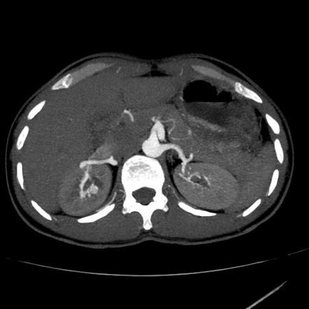
Renal Arteriovenous Malformation Radiology Reference Article Radiopaedia Org

The Borden Classification Of Dural Arteriovenous Fistulas Davf Groups These Lesions Into Three Types Based Upon The Site Of Ve Borden Classification Proposal

Amar Udare Md Radiogyan Com On Twitter Excellent Talk By Dshatzkes On Vascular Malformation At The Theapdr Noon Conference Take Home Point Be Careful Of What You Call A Hemangioma Refer

Pin On Snc

Vascular Malformation Classification References T M O Couto Download Scientific Diagram

Endovascular Treatment Of Arteriovenous Malformations Of The Head And Neck Focus On The Yakes Classification And Outcomes Journal Of Vascular And Interventional Radiology

Vascular Malformation Classification References T M O Couto Download Scientific Diagram

What Are Vascular Anomalies Ucsf Radiology

Pin By Marek Tupy On Education Ultrasound Physics Radiology Imaging Mri Brain

Pdf Classification Diagnosis And Interventional Radiologic Management Of Vascular Malformations Semantic Scholar
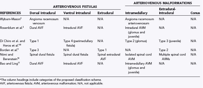
Classification Of Spinal Arteriovenous Lesions Neupsy Key

Pericallosal Lipoma Tubulonodular Radiology Case Radiopaedia Org Radiology Radiology Imaging Brain Images

Racing Car Sign Radiology Reference Article Radiopaedia Org Radiology Brain Images Medical Imaging

Primary Cns Lymphoma Radiology Case Radiopaedia Org Radiology Cns Radiology Imaging

Pulmonary Avm Radiology Case Radiopaedia Org Pulmonary Radiology Vascular

Yakes Avm Classification System 8 Download Table


Comments
Post a Comment